


- Mon - Thu:
- 08:30 - 12:00, 14:30 - 18:00
- Fri:
- 08:30 - 12:00
- Mon - Tue; Fri:
- 08:30 – 12:00
- Wed:
- 14:30 – 18:00
- Mon - Fri:
- 08:30 – 10:30
(Capacity limited – we ask that you register by telephone)
Services
- Dizziness questionnaire
- The patient is firstly asked to complete a questionnaire over several pages regarding his or her disorder. The questions are in relation to the properties of the dizziness symptoms, existing illnesses and medicines being taken.
- Specific discussion on dizziness
- After the dizziness questionnaire has been evaluated and all results provided by the patient have been reviewed, a detailed discussion with the patient is held in order to obtain further information on the illness.
- Standing and walking tests
- Standing and walking tests as developed by Unterberger and Romberg are among the easiest and at the same time most important test methods. These test methods were named after their inventors. They deliver important indications regarding dizziness disorders. In particular, they provide significant information on disruptions of the balance organ in the inner ear or disturbances in the area of the cerebellum.
- Electro or video nystagmography
- If there is a disturbance in the balance system, eye movements that are mostly involuntary occur. This condition is called nystagmus. The eye movements are analysed in a darkened room using infrared video glasses. The results are analysed automatically by a computer and may already provide initial indications on the cause of the illness, particularly in dizziness appearing acutely.
- Location and position testing using video oculography
- In this test, the patient’s eye movements are analysed using video-oculography computer recording with the patient's body in various positions. For example, if small calcium crystals (otoliths) have become lost in the ear, they may lead to movement-dependent dizziness (benign positional vertigo). This test can be used to verify the presence of this condition.
- Head Impulse Test (HIT) / Halmagyi test using video-oculography analysis
- One of the main functions of the balance system is to stabilise the field of view, i.e. in head or body movements, the eyes are always angled away from the direction of movement, so that the person always maintains a stable field of view even during a movement. This field of view stabilisation is enabled by what is referred to as the “vestibulo-ocular” reflex. Thanks to the latest technology, we are now in a position to test this reflex using video glasses that are extremely light and record very quickly. This enables an examination of all three movement axes in the area for the first time.
- Caloric testing using video-oculography analysis
- During this test, the most sensitive receptor of the balance organ, referred to as the horizontal arch, and the upper balance nerve are tested separately for each side using cold air.
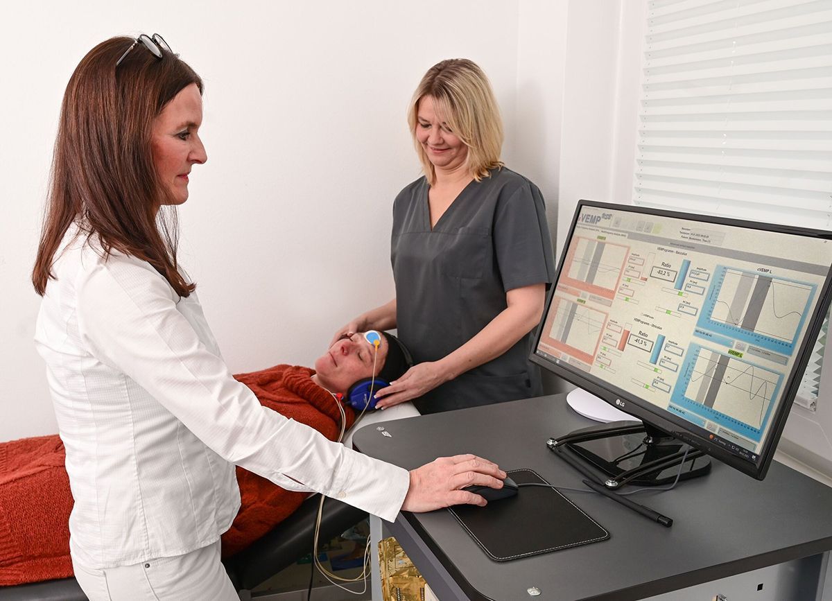
- Cervical vestibular evoked myogenic potentials (cVEMP, oVEMP)
-
The aforementioned head impulse test as well as video nystagmography measurement methods determine the capacity of the inner ear to perceive rotating movements. The measurement of vestibular evoked myogenic potentials (VEMPs) enables the functional inspection of the scacculus (c-VEMPs) and the utriculus (o-VEMPs) separately for each side.
The oVEMPs tests the utriculus, as well as the nervus vestibularis superior (superior balance nerve), for example. Among other things, the utriculus is responsible for horizontal movements. The measurement is used as a supplement to the VNG tests and is applied for the diagnosis of Superior Semicircular Canal Dehiscence (SSCD) or total vestibular failure, for example.
The vestibular-ocular reflex can be used in the oVEMP measurement. The potential difference of the extra-ocular musculature is measured using surface electrodes. In order to obtain good results, the patient looks straight up. The point of this is not to tighten the muscle more, but to bring the lower oblique muscle closer to the electrode.In the cVEMPs, the sacculus is tested, as well as the nervus vestibularis inferior (inferior balance nerve), for example. The sacculus is responsible for the perception of vertical movements. The measurement is used as a supplement to the VNG tests and is applied for the diagnosis of Superior Semicircular Canal Dehiscence (SSCD), Morbus Menière, partial or total vestibular failure/Neuritis Vestibularis, vestibular migraines, vestibular schwannoma or otosclerosis.
The vestibular-collic reflex is used in the cVEMP measurement. The patient flexes the M. sterocleidomastoideus by turning his or her head to the contra-lateral shoulder (turned away from the stimulus side). The contraction of the extraocular musculature triggered by the stimulus is measured ipsilaterally using surface electrodes. The tension reduces when the stimulus is supplied. The louder the stimulus, the greater the reflex. - Posturography and determination of risk of falling
- A second important function of the balance system is the maintenance of balance in the body. As the name indicates, posturography is used to test a patient's posture. In this test, the body's swaying motion is analysed within the scope of four different test situations (eyes open or closed, with or without foam cushion). Depending on the sway template, it is possible to draw conclusions on the cause of the dizziness condition and estimate the individual risk of falling.
- Determination of the subjective visual verticals
- By determining the subjective visual verticals, the functioning of the utriculus can be inspected during the differential diagnostics of the otolith apparatus in the balance organ. The otolith organs – utriculus and sacculus – in the balance labyrinth play a key role in the correct spatial orientation and stabilisation of the body's position. For a patient suffering from vertigo, the world is not upright. Information on this can be obtained by measuring the subjective visual verticals. To achieve this, a laser line has to be set is a straight position in a darkened room. The result provides the doctor with an insight into where the problem could lie early in the diagnostic process and is additionally useful for monitoring progress.
- 3D imaging (DVT)
- In dizziness diagnosis, alongside magnetic resource imaging (MRT) of the skull to assess the brain, digital volume tomography (in short: DVT) is also available for the precise diagnosis of the middle and inner ear structures as well as the upper cervical spine. In digital volume tomography, a spatial X-ray image of the skull is generated which can be observed in all levels and all layers. It is possible to view an individual point from various directions at the same time. The high imaging performance, which can be seen in both the maximum resolution and the effectively minimised radiation exposure, is a form of technical progress which directly benefits our patients.
Please understand that most of the services listed above may not be provided at the expense of the statutory health insurers. They have to be paid by the patient according to GOÄ digits (private fee schedule).
